NIVA Study Guide
National IV Association Exam Blueprint
Topic I. Veins and Arteries
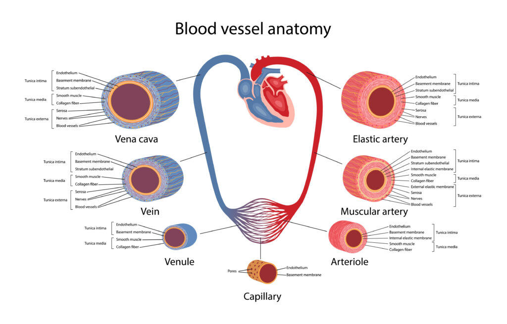
The walls of your veins are made up of three different layers:
- Tunica externa. This is the outer layer of the vein wall, and it’s also the thickest. It’s mostly made up of connective tissue. The tunica externa also contains tiny blood vessels called vasa vasorum that supply blood to the walls of your veins.
- Tunica media. The tunica media is the middle layer. It’s thin and contains a large amount of collagen. Collagen is one of the main components of connective tissue.
- Tunica intima. This is the innermost layer. It’s a single layer of endothelium cells and some connective tissue. This layer sometimes contains one-way valves, especially in the veins of your arms and legs. These valves prevent blood from flowing backward.
Veins are often categorized based on their location and any unique features or functions.
Pulmonary and systemic veins
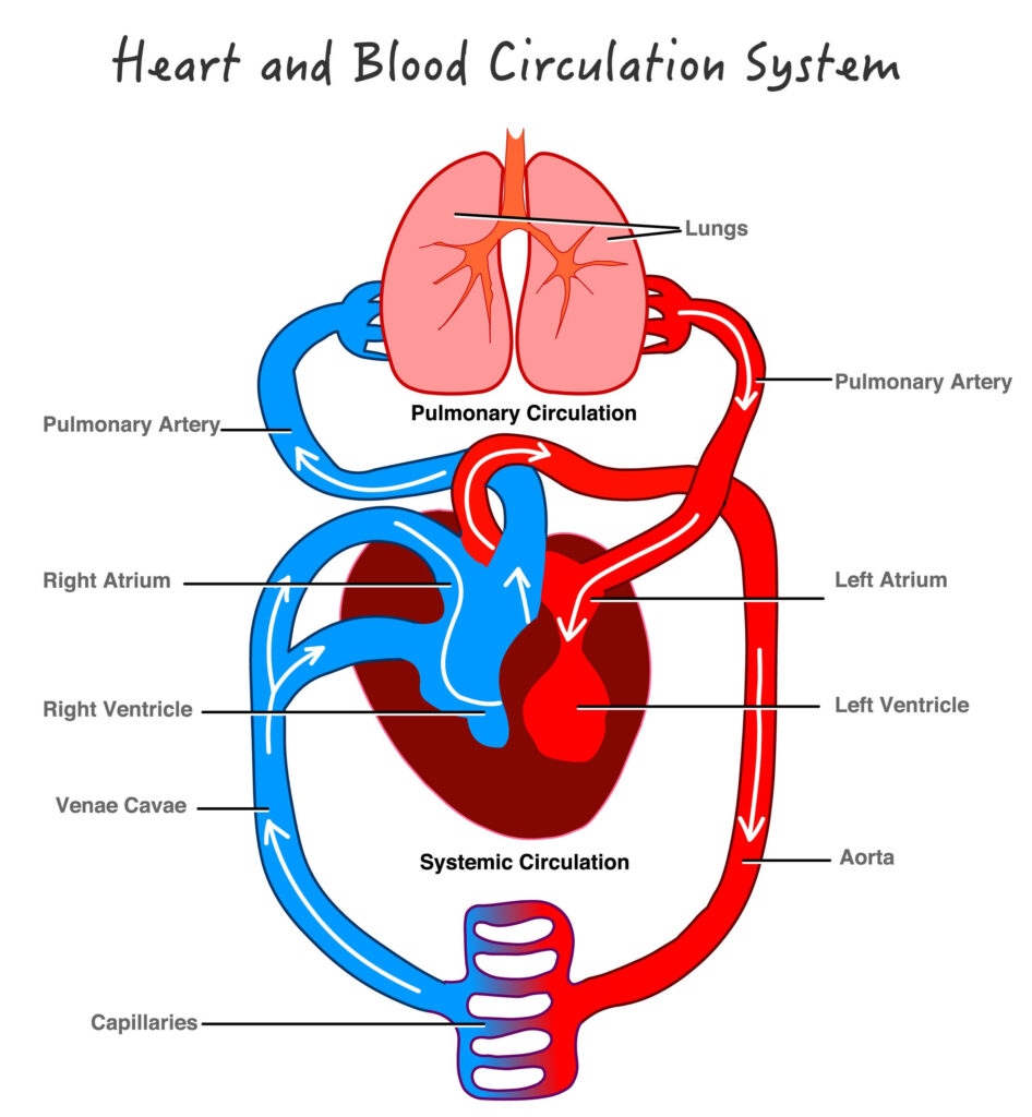
Your body circulates blood on two different tracks called the systemic circuit and the pulmonary circuit. Veins are based on the circuit they’re found in:
- Pulmonary veins. The pulmonary circuit carries deoxygenated blood from your heart to your lungs. Once your lungs oxygenate the blood, the pulmonary circuit brings it back to your heart. There are four pulmonary veins. They’re unique because they carry oxygenated blood. All other veins carry only deoxygenated blood.
- Systemic veins. The systemic circuit carries deoxygenated blood from the rest of the body back to your heart, where it then enters the pulmonary circuit for oxygen. Most veins are systemic veins.
Deep veins and superficial veins
Systemic veins are further classified as being either:
- Deep veins. These are found in muscles or along bones. The tunica intima of a deep vein usually has a one-way valve to prevent blood from flowing backward. Nearby muscles also compress the deep vein to keep blood moving forward.
- Superficial veins. These are located in the fatty layer under your skin. The tunica intima of a superficial vein can also have a one-way valve. However, without a nearby muscle for compression, they tend to move blood more slowly than deep veins do.
- Connecting veins. Blood from superficial veins is often directed into the deep veins through short veins called connecting veins. Valves in these veins allow blood to flow from the superficial veins to your deep veins, but not the other way.
Which conditions affect the venous system?
Many conditions can affect your venous system. Some of the most common ones include:
- Deep vein thrombosis (DVT). A blood clot forms in a deep vein, usually in your leg. This clot can potentially travel to your lungs, causing pulmonary embolism.
- Superficial thrombophlebitis. An inflamed superficial vein, usually in your leg, develops a blood clot. While the clot can occasionally travel to a deep vein, causing DVT, thrombophlebitis is generally less serious than DVT.
- Varicose veins. Superficial veins near the surface of the skin visibly swell. This happens when one-way valves break down or vein walls weaken, allowing blood to flow backward.
- Chronic venous insufficiency. Blood collects in the superficial and deep veins of your legs due to improper functioning of one-way valves. While similar to varicose veins, chronic venous insufficiency usually causes more symptoms, including coarse skin texture and ulcers in some cases.
What are the symptoms of a venous condition?
While the symptoms of a venous condition can vary widely, some common ones include:
- inflammation or swelling
- tenderness or pain
- veins that feel warm to the touch
- a burning or itching sensation
These symptoms are especially common in your legs. If you notice any of these and they don’t improve after a few days, make an appointment with your doctor.
They can perform a venography. In this procedure, your doctor injects contrast die into your veins to produce an X-ray image of a particular area.
Artery vs. vein
Arteries are blood vessels responsible for carrying oxygen-rich blood away from the heart to the body. Veins are blood vessels that carry blood low in oxygen from the body back to the heart for reoxygenation.
Arteries and veins are two of the body’s main type of blood vessels. These vessels are channels that distribute blood to the body. They’re part of two closed systems of tubes that begin and end at the heart. These systems of tubes are either:
- Pulmonary. The pulmonary vessels are arteries that transport oxygen-poor blood from the heart’s right ventricle to the lungs. Pulmonary veins transport oxygen-rich blood back to the heart’s left atrium.
- Systemic. The systemic vessels are arteries that carry oxygen-rich blood from the heart’s left ventricle to the tissues in all parts of the body. They then return oxygen-poor blood through the veins back to the heart’s right atrium.

What are the different types of arteries?
There are three types of arteries. Each type is composed of three coats: outer, middle, and inner.
- Elastic arteries are also called conducting arteries or conduit arteries. They have a thick middle layer so they can stretch in response to each pulse of the heart.
- Muscular (distributing) arteries are medium-sized. They draw blood from elastic arteries and branch into resistance vessels. These vessels include small arteries and arterioles.
- Arterioles are the smallest division of arteries that transport blood away from the heart. They direct blood into the capillary networks.
What are the different types of veins?
There are four types of veins:
- Deep veins are located within muscle tissue. They have a corresponding artery nearby.
- Superficial veins are closer to the skin’s surface. They don’t have corresponding arteries.
- Pulmonary veins transport blood that’s been filled with oxygen by the lungs to the heart. Each lung has two sets of pulmonary veins, a right and left one.
- Systemic veins are located throughout the body from the legs up to the neck, including the arms and trunk. They transport deoxygenated blood back to the heart.
The walls of veins and arteries are both made up of three layers:
- Outer. Tunica adventitia (tunica externa) is the outer layer of a blood vessel, including arteries and veins. It’s mostly composed of collagen and elastic fibers. These fibers enable the veins and arteries to stretch a limited amount. They stretch enough to be flexible while maintaining stability under the pressure of blood flow.
- Middle. The middle layer of the walls of arteries and veins is called the tunica media. It’s made of smooth muscle and elastic fibers. This layer is thicker in arteries and thinner in veins.
- Inner. The inner layer of the blood vessel wall is called tunica intima. This layer is made of elastic fiber and collagen. Its consistency varies based on the type of blood vessel.
Unlike arteries, veins contain valves. Veins need valves to keep the blood flowing toward the heart. These valves are particularly important in the legs and arms. They fight gravity to prevent the backflow of blood.
Arteries don’t need valves because the pressure from the heart keeps the blood flowing through them in one direction.
The cardiovascular system is a closed system of vessels called arteries, veins, and capillaries. They’re all connected to a muscular pump called the heart. The cardiovascular system keeps a continuous and controlled movement of blood that delivers nutrients and oxygen to every cell in the body. It does this through thousands of miles of capillaries between the arteries and the veins.
- Arteries. The pulmonary arteries carry low-oxygen blood from the right ventricle of the heart to the lungs. Systemic arteries transport oxygenated blood from the left ventricle of the heart to the rest of the body.
- Veins. The pulmonary veins carry oxygenated blood from the lungs to the left atrium of the heart. Systemic veins carry low-oxygen blood from the body to the right atrium of the heart.
- Capillaries. Capillaries are the smallest and most numerous of the blood vessels. They connect the arteries (which carry blood away from the heart) and the veins (which return blood to the heart). The primary function of capillaries is the exchanging of materials, like oxygen, between the blood and tissue cells.
- Heart. The heart has four chambers: the right atrium, right ventricle, left atrium, and left ventricle. The heart provides the force to circulate blood through the cardiovascular system.
Nutrients and oxygen are delivered to every cell in your body through a circulatory system. The heart pumps oxygenated blood to your cells through arteries. It pumps oxygen-depleted blood away from your cells through veins.