Disclaimer: This course and certification does not give the student any authority whatsoever to start and IV or begin IV access. This course does not license anyone to start IV.’s. This course does not give or elude to give authority which it has none to students taking this course to flush, or hang medication.
Flushing an IV , pushing medication, hanging medication must only be done by licensed professionals which are RN’s or Physicians.
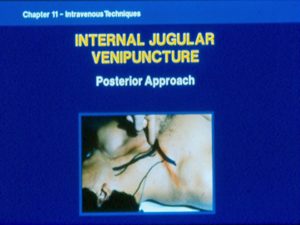
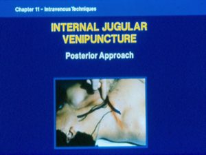
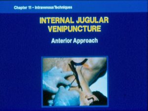
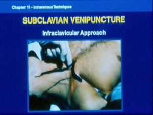
IV Part I of 5
IV Part 2 of 5
IV Part 3 of 5
IV Part 4 of 5
IV Part 5 of 5
This course does prepare the student to have a deeper understanding of IV.s, the reasons of necessity, the complications associated with IV insertions and the correct means and techniques of insertion, provide an explanation of fluid balance and sign and symptoms associated with those patient’s who may be in need of an IV.
This course is designed to help RN’s, LPN’s and other licensed and non-licensed personal to become more capable at starting an IV, finding veins and the like
For the layperson to start IV access they must be working in an environment which covers them with the liability do preform this task or be working under a physician’s license.
Peripheral intravenous (IV) device management
Introduction
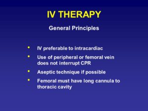
Peripheral intravenous device (PIV)/catheters are the most commonly used intravenous device in hospitalized patients. They are primarily used for therapeutic purposes such as administration of medications, fluids and/or blood products as well as blood sampling. PIV’s are usually considered a low risk, however it can be associated with complications such as hematoma, phlebitis, pain and infections.
Aim
The aim of this guideline is to provide an outline of insertion and securing of the PIV line and ongoing management of the PIV device including infection control in hospital, outpatient, and home healthcare settings and management of common complications.
Definition of terms
Peripheral IV devices: are cannula/catheter inserted into a small peripheral vein for therapeutic purposes such as administration of medications, fluids and/or blood products.
Aseptic technique: aims to prevent pathogenic microorganisms in sufficient quantity to cause infection, from being introduced to susceptible sites by hands, surfaces and equipment.
Decontaminate hands: Perform hand hygiene (moment 2) – to protect the patient from harmful germs from entering their body during a procedure.
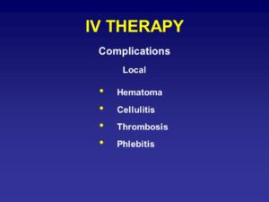
Phlebitis: swelling, redness, heat, and pain related to local inflammation of the vein at or near the cannula site.
Infiltration: occurs when fluid infuses into the tissues surrounding the venipuncture site. This sometimes happens when the tip of the catheter slips out of the vein, the catheter passes through the wall of the vein, or the blood vessel wall allows part of the fluid to infuse into the surrounding tissue
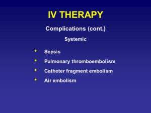
Extravasation: An extravasation occurs when there is accidental infiltration of a vesicant or chemotherapeutic drug into the surrounding IV site.
Haematoma: Hematoma is a mass of blood confined to a space in tissue or organ due to a break in a blood vessel.
Pump: refers to infusions pumps eg. Large volume pumps (LVPs)/Volumetric pumps e.g Alaris Signature Edition (SE), Syringe drivers (e.g. Alaris GH+), Patient Controlled Analgesia/PCA pumps (Alaris PCAM), Ambulatory Pumps (CME Niki T34, Smiths Medical CADD pump).
Double checking: refers to two clinicians (appropriately endorsed Enrolled nurses (EN), Registered Nurses (RN), Doctors or Pharmacists) independently checking the medications.
Assessment
Assessment during PIV maintenance: Both local and systemic assessments should be completed.
Assessing the IV equipment, PIV infusion site, and holistic assessment of the patient should be done on a regular basis.
In an inpatient setting, six hourly assessments are a minimum if the child is not on continuous infusion. Unstable patients who have signs and symptoms of complications would be assessed more frequently.
If the patient is receiving continuous IV infusion, observations of the IV site, rate of infusion and fluid checks are observed hourly and documented in the fluid balance flowsheet. Once documented review the overall fluid status of the child in the fluid balance activity .
PIV cannula are considered a high risk for pressure injury. As per Pressure Injury prevention guideline, the PIV site should be checked hourly for pressure sore and any signs of infection unless documented otherwise.
For HITH patients, the nurse will assess the PIV with each visit. Education will be provided to parents on the signs of pressure injuries and the process of contacting the HITH nurse.
Management
General considerations for PIV insertion:
Use aseptic technique when preparing and administering fluids and medications
Adhere to six rights of medication safety
Prepare patient and family for the procedure
Involve play and distraction techniques, relaxation and other coping skills appropriate to the age of the child see Comfort Kids techniques.
Consider nitrous oxide for anxious children
Consider children who have prior traumatic experience with IV insertion
Acute management
Pain Management
Infants <3 months:
Oral sucrose with a pacifier should be used (see Procedure management guideline) or encourage mother to feed infant during IV insertion procedure.
Give parent or care giver option to hold infant during IV insertion procedure & employ multisensory stimulation
Older infants & children:
Minimum 45-60 minutes prior to cannulation apply local anesthetic cream (e.g EMLATM -60 mins, AnGelTM – 45 mins) to several possible IV sites and cover with transparent film dressing. http://www.rch.org.au/rchcpg/hospital_clinical_guideline_index/Procedural_Pain_Management
Use distraction techniques prior to and during the procedure see Comfort Kids techniques. Prepare appropriate methods of distraction for the child. Ascertain from the child and family what techniques are most likely to attract their attention. For example, pop-up books, musical books, blowing bubbles and guided imagery. Use of Buzzy while inserting IV cannula to distract the child. http://buzzy4shots.com/
Securing of IV Cannula
Dressings to PIV sites are the first line of defence against infections and must be kept secure, clean and dry.
The type of secure dressing for the PIV cannula depends upon the child’s age, condition of the skin, site of the IV, child’s activity and/or or mobility.
Consider placing a small piece of cotton wool ball or gauze underneath the hub of the cannula to reduce pressure.
Cover the cannula site with sterile transparent semipermeable occlusive dressing (e.g. Tegaderm, IV 3000) placed aseptically over the catheter.
If desired, place sterile tape over the hub and wings of the device before placing the transparent dressing.
IV board / splints are recommended to secure PIV cannula placed in or adjacent to areas of flexion. This will adequately immobilise the joint and minimise the risk of venous damage resulting from flexion.
When using Splints, ensure these are positioned and strapped with the limb and digits in a neutral position to prevent restricting blood or nerve supply and pressure sores.
Inspect the splint at least daily and change if soiled by blood or fluid leakage.
Cover with gauze or non-compression tubular bandage
When using non compression tubular bandage (e.g. Tubifast), ensure there is a clear window where the cannula enters the skin so the site can be viewed
In Summary, when dressing a peripheral IV cannula ensure:
it is secure,
the site is visible
the child can’t injure themselves on the connections
the child can’t remove or dislodge the cannula
that tapes are not too tight.
Refer to Intravenous access – Peripheral guideline for the procedure and steps involved in securing the cannula
Change the dressing only if it becomes insecure or if there is blood or fluid leakage.
PIV dressing
Ongoing management
Administration of Bolus/loading doses
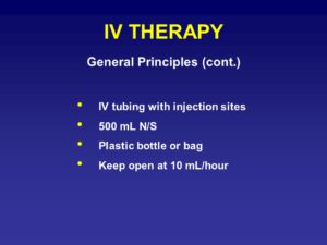
If the cannula is accessed infrequently for the administration of medications or fluids, it should be flushed with Normal saline only as evidence suggests equal or no benefit of using hepsaline. The cannula should be flushed on a 6 hrly basis if accessed intermittently.
For Opioid infusion bolus refer to the specific guidelines: Children’s Pain Management Service (CPMS)(opioid infusion guideline)
Administration of intravenous fluid, drug infusions or Blood products through IV device
Monitoring and evaluation of patients on fluid therapy is mandatory to prevent the occurrence of fluid overload.
Assessments are completed hourly to determine that the fluid infusing is as per medical prescription. Check the solution is the prescribed one, the rate of infusion, the amount infused and the remaining amount to be infused is confirmed as per the order in the Medical Administration Record (MAR).
Drugs administered as an IV infusion may be inserted into a bag of IV fluids, the burette of an infusion set for administration via a volumetric infusion pump or in a syringe for use in a syringe driver. The most appropriate method should be selected depending on volume of diluent required and intended rate of delivery.
Drugs administered via:
Burette of an infusion set: to dilute the drug in a smaller volume via burette giving system- hang the bag of infusion fluid and gradually open the roller camp to allow appropriate amount of diluent into the burette. Inject the prescribed drug into the burette via the additive port. Ensure the drug is well mixed in burette by shaking the burette.
Syringe driver: is recommended for children weighing less than 10 kg. Draw up required volume of diluent in appropriate size syringe and then pull back the syringe plunger to enable you to inject the drug into the syringe using aseptic technique. Ensure the drug is well mixed in the syringe by gentle shaking.
Infusion bag: Clean the rubber bung with alcohol swab before injecting prepared drug into infusion fluid bag via the additive port and mix well by shaking the bag. Without contaminating the key parts insert the spike on the administration set into the septum of the infusion bag.
Attach a completed drug label detailing the drug, dose, diluent, volume of diluent, date, time and signature of the nurse and the staff who double checked
Access the IV cannula only after cleaning the rubber bung with alcohol swab (Scrub the hub)
For intermittent infusions, IV lines can be disconnected between infusions, but ensure the cannula is flushed with appropriate flush once IV line is disconnected from the cannula.
Administering blood products
Check patient and blood product identification as per the Blood Product Transfusion Procedure.
Administer blood product transfusions via a volumetric infusion pump or syringe driver to ensure accurate delivery. Use gravity sets only when rapid administration is required with diligent monitoring of volume.
Use a Neonatal transfusion set (includes a 170 to 200 micron filter required for blood products) and syringe driver for delivering small volumes of blood products.
Using aseptic technique, spike the blood product with the Neonatal transfusion set and attach an appropriate sized syringe for the transfusion to the 3 way tap.
Draw the required volume into the syringe and prime the rest of the neonatal transfusion set. Label the syringe with both patient and blood product identification details including expiry date and time of blood product.
If rapid transfusion of small volumes is required, draw the required volume into a syringe through a 170 to 200 micron filter.
Burettes should not be used for transfusion of blood products
What Fluids and how much fluids to use
Refer to the Intravenous Fluids Clinical Practice Guideline: Intravenous Fluids
PIV table
Changing cannulas
Re-cannulation should be avoided where possible, as this will cause the child and family further distress. There is no limit to the length of time that a cannula may remain in situ and with appropriate care, several days may be possible. Cannulas only need to be replaced when there is accidental dislodgement, occlusion, Phlebitis and infection.
Removal of IVs
The possible reasons for removal of cannula includes: Infiltration, extravasation, no longer be required, no longer be functioning effectively or it may be causing the child excessive discomfort, signs of phlebitis or infection.
Perform hand hygiene, wearing non-sterile gloves, carefully remove the dressing, holding the cannula in place at all times.
Hold a piece of sterile gauze or cotton wool over the exit site but do not apply pressure
Slowly withdraw the cannula, maintaining a neutral angel with the child’s skin.
Cover site with cotton wool and tape or Band-Aid.
Advise the child and family that the cotton wool and tape or Band-Aid should remain in situ for 24 hrs.
Document date and reason of removal.
Prevention of infections
Good hand hygiene before PIV catheter insertion and maintenance, combined with proper aseptic technique during catheter manipulation provides protection against infection.
Prior to accessing PIV cannula, clean with an approved antiseptic wipe.
All children with a capped PIV access device in situ should have the site inspected at commencement of shift and least every six hours for signs of infusion phlebitis.
Where children are receiving continuous IV fluids/medication, site should be inspected hourly and documented on the fluid balance flowsheet for the duration of the infusion.
Management of complications
Complications associated with IV therapy are common. Most are preventable by attention to IV infusion equipment, aseptic technique and attention to fluid and electrolyte prescribing. Common problems are
Infection:
Skin-based bacteria may enter through insertion site
Local cellulitis or systemic bacteraemia are possible.
If infection is present, remove the IV cannula immediately, swab the insertion site and contact medical team to review.
Phlebitis: Vein irritation
due to the presence of the catheter/fluids or medication
Chronically ill patients requiring multiple and recurrent IV access.
Notify medical team to review and document in patient record
Infiltration: occurs when fluids or medications leaks into surrounding tissue. If infiltration occurs:
Immediately stop the infusion and disconnect the tubing as close to the catheter hub as possible.
Remove the catheter without placing pressure on the site.
Elevate the affected limb.
Continue to assess and document the appearance of the site and associated signs and symptoms.
Extravasation: delivery of fluids or medications into surrounding tissue.
If extravasation occurs, assess the Grade of extravasation Injury. (Refer neonatal extravasation guideline).
Most extravasation injuries are of Grades 1 & 2 and do not require extensive intervention to prevent long-term skin and soft tissue damage.
Grade 3 & 4 injuries have a greater potential for skin necrosis, compartment syndrome and need for future plastic surgery, depending on the type of solution extravasated. (Refer neonatal extravasation guideline).
Other considerations:
The infant’s parents should be informed of an extravasation injury and management plan.
An incident report should be completed for grade 3 & 4 extravasations.
Documentation
Care of peripheral IVs
Peripheral IV site – document the presence of any atypical findings or complications and any actions taken in the LDA (Lines, Drains and Airway) section of EMR/progress notes. VHIMS should be completed for all IV extravasations.
Fluids and medications through peripheral IV’s
Pump pressures for each IV line: should be documented hourly on Fluid balance activity. The blue Alaris pumps have a maximum pressure set and the pressure should not exceed 100mmHg
Infused volume: Hourly on fluid balance flowsheet (it is advised to clear the infusion pump hourly.)
Any complications
Record the date and time of the infusion when extravasation was noted, the type and size of catheter, the drug administered, the estimated amount of extravasated solution, and the administration technique used.
Record the patient’s signs and symptoms, treatment, and response to treatment. Include the time you notified the patient’s primary care provider and the primary care provider’s name.
Unit 1: Legal Aspects of who can start an IV


Micr
IV test This is a great practice test. You may print this out. If you would like it graded, please contact your instructor.
Please click on each link below and read its contents before continuing.
PeripheralIntravenousInitiationModule
________________________________________________________________________________________________________________________________________________
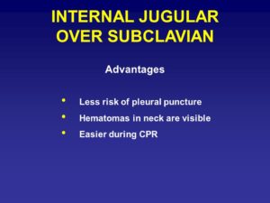
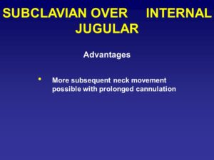
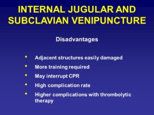
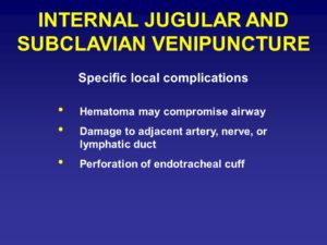
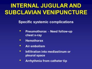
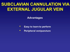
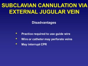
Unit 2: Complications associated with intravenus insertion
Unit 3: IV fluids