Estimated reading time: 15 minutes
Intravenous (IV) therapy is one of the most commonly performed invasive procedures in healthcare. IV access is used for fluid resuscitation, medication administration, chemotherapy, immunosuppressant therapy, vitamin and micronutrient therapy, blood collection, and emergency access.
Because an IV catheter provides direct access to the vascular system, complications can sometimes progress quickly. Problems such as phlebitis, infiltration, extravasation, catheter-related infection, occlusion, thrombosis, air embolism, and fluid overload require prompt recognition and appropriate intervention.
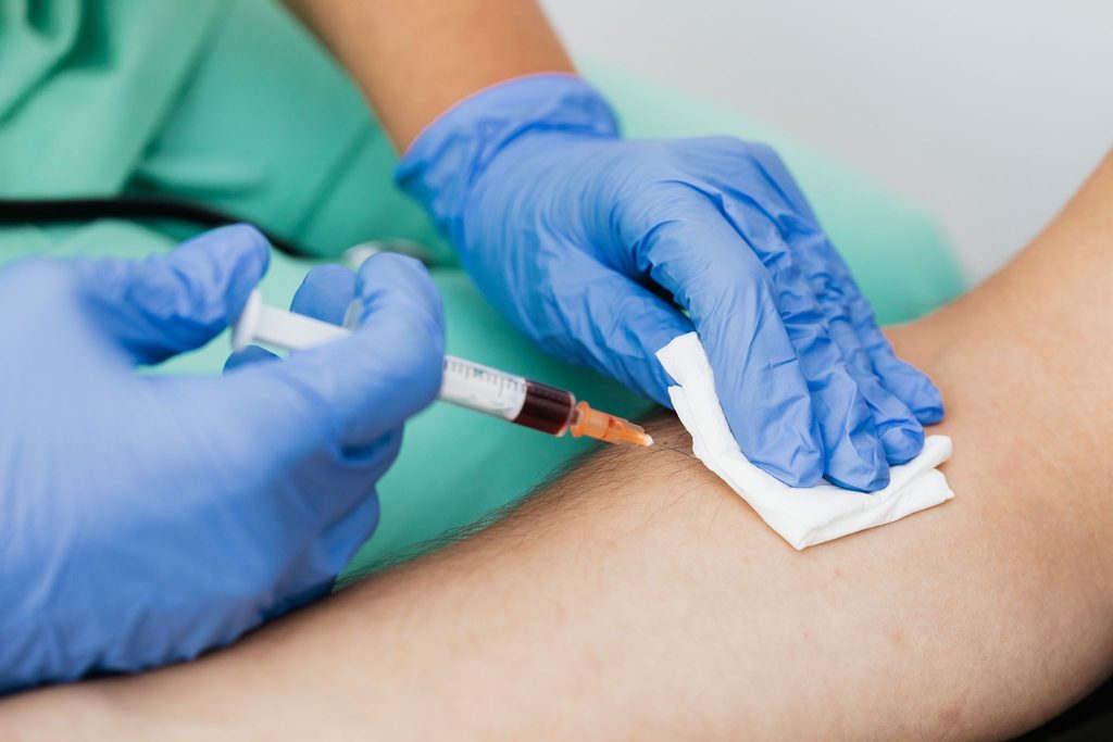
For student nurses, nurses, anesthesia techs, veterinary techs, dentists, and healthcare educators who teach IV skills, understanding the warning signs of IV complications is an essential part of safe clinical practice.
This guide explains how to recognize common IV complications, understand their causes and risk factors, respond appropriately, document events, and teach IV complication prevention through clinical education and simulation.
Table of contents
- What Are the Most Common IV Complications?
- Why IV Complication Management Matters
- Understanding the Anatomy Behind IV Complications
- Choosing the Right Vein Can Help Prevent Complications
- IV Insertion Technique and Complication Prevention
- Sterile Technique and Skin Antisepsis
- Understanding IV Solutions and Their Effects
- Fluid Overload and Rapid Infusion
- Phlebitis: Signs, Causes, and Management
- Infiltration: Recognition and Management
- Extravasation: A More Serious Tissue Injury Risk
- Catheter-Related Infection
- Catheter Occlusion and Thrombosis
- Air Embolism: Rare but Serious
- Phlebitis and Infiltration Assessment Scales
- IV Flushing and Blood Draws
- Medication Verification and IV Safety
- IV Documentation: What Should Be Charted?
- Scope of Practice and Professional Accountability
- How IV Instructors Can Teach Complication Recognition
- Applying Erikson’s Psychosocial Stages to IV Education
- Geriatric IV Complication Considerations
- Education and Competency Assessment
- Interprofessional IV Responsibilities
- Preventing IV Complications at the System Level
- IV Safety Checklist
- Three IV Complication Scenarios to Recognize
- Final Practice Points for IV Clinicians and Educators
- Continuing Education and Professional Development
- Conclusion
What Are the Most Common IV Complications?
The most commonly encountered IV complications discussed in this guide include:
- Phlebitis — inflammation of the vein
- Infiltration — leakage of a non-vesicant IV solution into surrounding tissue
- Extravasation — leakage of a vesicant medication into surrounding tissue
- Catheter-related infection
- Catheter occlusion or thrombosis
- Air embolism
- Fluid overload
Early recognition is important because some IV complications remain localized while others can produce systemic effects.
Quick Answer: If an IV site develops unexpected pain, redness, swelling, coolness, blanching, resistance to flushing, loss of blood return, or other concerning changes, stop and assess the infusion according to applicable clinical protocols. The appropriate response depends on the suspected complication, medication or solution involved, patient condition, and institutional policy.
Why IV Complication Management Matters
IV therapy can produce rapid physiological effects because fluids and medications enter the vascular system directly.
Examples of potential problems include:
- Hypertonic solutions causing vascular irritation or fluid shifts
- Rapid infusions contributing to cardiovascular overload in vulnerable patients
- Breaks in aseptic technique contributing to catheter-related bloodstream infection
- Vesicant medications causing tissue injury when they escape the vein
Maintaining appropriate technique, verifying medication and fluid labels, following applicable authorization requirements, and documenting care are therefore fundamental components of IV safety.
Understanding the Anatomy Behind IV Complications
An IV catheter passes through several layers before entering the vein.
From the skin surface toward the vascular lumen, these include:
- Epidermis
- Dermis
- Hypodermis/subcutaneous tissue
- Venous wall, including the tunica intima, media, and adventitia
Understanding these structures helps clinicians understand the physical sensations associated with insertion, recognize flashback, and understand how fluid or medication can enter surrounding tissue if the catheter is not positioned appropriately.
Choosing the Right Vein Can Help Prevent Complications
Vein selection is an important part of IV complication prevention.
For peripheral IV access, commonly used veins include:
- Cephalic vein
- Basilic vein
- Median cubital vein
The antecubital fossa contains several relatively large veins that may be useful for peripheral access.
When selecting a vein, clinicians should:
- Trace the vein before insertion
- Identify a sufficiently straight segment
- Avoid areas over joints when possible
- Consider vessel quality
- Consider the patient’s condition and history
- Select an appropriate catheter size
The source recommends tracing the vein for at least an inch (approximately 2–3 cm) to understand its direction and continuity before cannulation.
Special Populations
Vein selection becomes particularly important in:
- Older adults with fragile or tortuous veins
- Pediatric patients with smaller vessels
- Patients with diabetes
- Patients with peripheral vascular disease
- Patients with cardiac or renal conditions affecting fluid management
Technique should always be adapted to the individual patient’s vessel quality and clinical condition.
IV Insertion Technique and Complication Prevention
Technical precision during IV insertion can help reduce mechanical injury.
Important principles include:
Insert at the Appropriate Angle
The source describes a peripheral IV insertion angle of approximately 15–28 degrees, depending on vein depth.
Keep the Bevel Up
The needle should be inserted with the bevel facing upward.
Recognize Flashback
Blood return in the catheter hub indicates entry into the vein. Once flashback is observed, the catheter should be advanced appropriately while reducing the angle to help avoid passing through the vessel.
Avoid Transfixing the Vein
Continuing to advance after entering the vein can cause the needle or catheter to pass through the posterior wall.
Never Force a Flush
Resistance during flushing can indicate:
- Malposition
- Catheter occlusion
- Placement against the vessel wall
- Perivascular placement
If resistance occurs, stop and reassess rather than forcing the flush.
Sterile Technique and Skin Antisepsis
Aseptic technique is fundamental to preventing infection and other IV complications.
Important practices include:
- Hand hygiene before and after the procedure
- Appropriate antiseptic skin preparation
- Allowing the antiseptic to dry completely
- Appropriate gloves
- Appropriate dressings and securement
- Maintaining a clean field during dressing changes and line access
The source identifies chlorhexidine as the preferred skin antiseptic for most adult patients and discusses cleansing the insertion site for approximately 30 seconds to one minute, followed by complete drying.
Because antiseptic products, concentrations, application times, and patient-specific precautions can vary, clinicians should follow the applicable current institutional protocol.
Understanding IV Solutions and Their Effects
Understanding solution tonicity is another important component of IV education.
| Solution | General Effect | Clinical Considerations |
| 0.9% Normal Saline | Isotonic and closely aligned with extracellular fluid | Used for applications such as resuscitation and medication dilution; large volumes may contribute to hyperchloremic metabolic acidosis in some patients |
| 3% Hypertonic Saline | Draws water toward the vascular space | Used in selected situations such as severe hyponatremia and cerebral edema; requires careful administration |
| 0.45% Saline | Hypotonic; shifts fluid toward cells/interstitium | Used in selected cases of intracellular dehydration; inappropriate use can contribute to complications |
Medication and fluid labels should always be carefully checked. Confusing solutions with medications such as atropine or lidocaine can result in serious medication errors.
Fluid Overload and Rapid Infusion
Infusion speed must be appropriate for the patient’s physiology.
Patients who may be particularly vulnerable to cardiovascular overload include some:
- Older adults
- Patients with heart failure
- Patients with renal impairment
- Other medically fragile patients
Potential warning signs include:
- Tachycardia
- Elevated blood pressure
- Shortness of breath
- Tachypnea
- Low oxygen saturation
- Jugular venous distension
- Crackles on lung auscultation
When signs of acute overload occur, the source recommends stopping the infusion, positioning the patient upright, administering oxygen, and notifying the provider, while following applicable emergency protocols.
Phlebitis: Signs, Causes, and Management
What Is Phlebitis?
Phlebitis is inflammation of the vein wall.
It can be:
- Chemical — associated with an irritating medication or solution
- Mechanical — associated with catheter movement or irritation
- Infectious — associated with infection
Common signs include:
- Redness along the vein
- Pain or tenderness
- Warmth
- Swelling
- A palpable or tender cord
- Possible purulent drainage when infection is involved
What Should You Do if Phlebitis Is Suspected?
The source recommends:
- Stop the infusion.
- Discontinue the catheter when phlebitis is suspected.
- Assess for systemic signs of infection.
- Establish alternative access when necessary.
- Obtain cultures or notify the prescriber when infection is suspected.
- Document the event thoroughly.
Documentation should include the time, catheter information, site, signs and symptoms, patient response, and actions taken.
Infiltration: Recognition and Management
What Is Infiltration?
Infiltration occurs when a non-vesicant IV solution leaks into the surrounding tissue.
This differs from extravasation, where a vesicant medication escapes into tissue and can cause cellular injury.
Signs of Infiltration
Watch for:
- Coolness around the IV site
- Blanching
- Swelling
- Tightness
- Loss of blood return
- Resistance during flushing
What Should You Do?
If infiltration is suspected:
- Stop the infusion.
- Clamp or remove the catheter as appropriate.
- Elevate the affected limb.
- Follow the applicable compress protocol.
- Assess the affected area.
- Document the size, appearance, symptoms, and progression.
If the solution or medication involved requires an extravasation protocol, follow that protocol rather than treating it as ordinary infiltration.
Extravasation: A More Serious Tissue Injury Risk
What Is Extravasation?
Extravasation is the leakage of a vesicant medication into surrounding tissue.
Some chemotherapy agents and concentrated vasopressors are examples of agents that can cause significant tissue injury if extravasated.
Potential findings include:
- Severe pain
- Blanching
- Swelling
- Tissue injury
- Blistering
- Potential necrosis
Immediate Response
When extravasation is suspected:
- Stop the infusion immediately.
- Do not automatically remove the catheter.
- Follow the institution’s extravasation protocol.
- Preserve the catheter when required for aspiration.
- Consult the appropriate prescribing clinician and pharmacy.
- Follow medication-specific antidote and compress instructions.
- Document the event in detail.
Documentation should include the medication, dose, time, estimated amount involved, physical findings, and interventions.
Catheter-Related Infection
IV-related infection can occur locally or involve the bloodstream.
Local Signs
- Redness
- Tenderness
- Purulent drainage
Systemic Signs
- Fever
- Chills
- Hypotension
- Unexplained leukocytosis
When infection is suspected or confirmed, management should follow institutional and provider-directed protocols. This may include catheter removal, cultures, antimicrobial therapy, and infection-prevention measures.
Catheter Occlusion and Thrombosis
A catheter that becomes difficult or impossible to flush requires assessment.
Possible signs include:
- Inability to flush
- Reduced infusion flow
- Swelling
Potential causes include:
- Mechanical obstruction
- Fibrin sheath
- Thrombus
- Catheter positioning problems
Key Safety Rule
Do not forcibly flush against resistance.
First assess for mechanical or positional causes. Pharmacologic treatment such as a thrombolytic may be appropriate in selected circumstances and according to applicable protocols.
Air Embolism: Rare but Serious
Air embolism occurs when air enters the venous circulation.
Potential signs include:
- Sudden chest pain
- Dyspnea
- Hypotension
- Cyanosis
- Altered mental status
- Cardiac arrhythmia
Prevention
Prevention includes:
- Properly priming tubing
- Clamping lines appropriately
- Using occlusive dressings
- Preventing air entry during line insertion and removal
- Maintaining appropriate line connections
If an air embolism is suspected, follow the institution’s emergency-response protocol immediately. The source describes positioning the patient in left lateral decubitus and Trendelenburg when tolerated, administering 100% oxygen, and activating emergency response.
Phlebitis and Infiltration Assessment Scales
Standardized assessment tools can help clinicians consistently document changes at an IV site.
The source describes commonly used:
- Phlebitis scales
- Infiltration scales
Both use graded findings to help characterize severity.
Example Phlebitis Scale
| Grade | Findings | Example Action |
| 0 | No symptoms | Continue routine care |
| 1 | Possible tenderness/slight erythema | Monitor closely |
| 2 | Redness, swelling, pain along vein | Remove catheter and follow protocol |
| 3–4 | Palpable cord, marked erythema, possible purulence | Remove catheter; culture/treat when indicated |
Standardized assessment supports clearer documentation and communication between clinicians.
IV Flushing and Blood Draws
Flushing and blood collection from IV lines require careful attention to technique and timing.
Important considerations include:
- Use the appropriate saline and technique.
- Follow applicable facility requirements for flushing.
- Consider medication contamination when collecting blood from an IV line.
- Observe required timing between medication administration and blood collection.
- Follow policy regarding discard/waste volumes.
- Avoid placing a tourniquet above an arm with an active IV infusion.
The timing of blood collection can affect laboratory results when medications are being administered through the line, so institution-specific policies should determine the appropriate procedure.
Medication Verification and IV Safety
Medication errors can create serious IV-related complications.
Before administering an IV medication, verify:
- Patient
- Medication
- Concentration
- Dose
- Route
- Time
- Expiration date
- Applicable order
High-risk medications may also require two-person verification according to institutional policy.
Documentation should include relevant medication information, including lot numbers and administration times when required for traceability.
IV Documentation: What Should Be Charted?
Documentation should create a clear record of both routine IV care and complications.
Important information includes:
- Date and time
- Catheter gauge
- Catheter length
- Manufacturer when required
- Exact anatomical location
- Number of attempts
- Assistance provided
- Patient response
- Relevant vital-sign changes
- Interventions
- Patient/family education
- Provider notification
- Orders received
For students and novice clinicians, learning precise documentation is part of developing safe clinical practice.
Scope of Practice and Professional Accountability
IV skills must always be performed within the individual’s professional scope of practice.
Scope can depend on:
- Profession
- State regulations
- Employer policies
- Delegation
- Supervision
- Clinical setting
Medical assistants, for example, should consult supervising clinicians when IV responsibilities exceed their authorized scope.
For students and novice clinicians, supervision and progressively increased responsibility help support safe learning.
How IV Instructors Can Teach Complication Recognition
Effective IV education should combine knowledge with hands-on practice.
A strong skills program can incorporate:
Preparation
Provide:
- Appropriate equipment
- Simulation models
- Documentation templates
- Skills checklists
- Complication scenarios
Demonstration and Return Demonstration
Instructors should demonstrate the skill and then have learners perform it while being observed.
Scenario-Based Simulation
Simulation can recreate situations such as:
- Infiltration
- Extravasation
- Phlebitis
- Fluid overload
- Difficult access
- Medication-related complications
Constructive Feedback
Learners need opportunities to understand:
- What happened
- Why it happened
- What warning signs were present
- What could have been done differently
- How to respond safely next time
The source emphasizes preparation, passion, patience, individualized coaching, debriefing, and continuous learning as important instructor practices.
Applying Erikson’s Psychosocial Stages to IV Education
IV education is not only about technical skill.
Patients and learners have different developmental, emotional, and communication needs.
Erikson’s psychosocial framework can help instructors adapt communication across the lifespan.
Examples include:
- Infants: reduce sensory distress and involve caregivers appropriately.
- Toddlers: offer simple choices.
- Preschool children: use play and age-appropriate explanations.
- School-age children: encourage participation and competence.
- Adolescents: respect privacy and autonomy.
- Young adults: encourage collaborative decision-making.
- Middle adults: connect care with health and family responsibilities.
- Older adults: emphasize dignity, autonomy, life experience, and individual goals.
Using developmentally appropriate communication can make IV education more patient-centered and responsive.
Geriatric IV Complication Considerations
Older adults may have characteristics that require additional attention during IV therapy.
These can include:
- Fragile veins
- Thinner skin
- Reduced cardiac reserve
- Altered fluid distribution
- Polypharmacy
For older patients, instructors should emphasize:
- Appropriate catheter selection
- Gentle securement
- Protection of fragile skin
- Careful fluid monitoring
- Medication review
- Clear and respectful communication
- Appropriate caregiver involvement
Fluid overload may present subtly, so learners should understand the importance of monitoring the patient’s overall clinical condition rather than relying on a single measurement.
Education and Competency Assessment
A structured IV competency program can include four major components:
1. Didactic Education
Cover:
- Anatomy
- Infection prevention
- IV solutions
- Medication safety
- Complication recognition
2. Simulation
Use task trainers and simulated complications to practice recognition and response.
3. Supervised Clinical Practice
Learners should perform procedures under appropriate supervision and receive feedback.
4. Documentation Assessment
Evaluate whether learners can accurately document:
- Time
- Gauge
- Location
- Attempts
- Patient response
- Complications
- Interventions
Interprofessional IV Responsibilities
IV care often involves multiple members of a healthcare team.
The source highlights different educational considerations for:
Student nurses and new nurses:
Perform IV procedures under appropriate supervision until competency is established.
Anesthesia technicians:
Understand IV monitoring, line setup, and emergency response in procedural environments.
Veterinary technicians:
Adapt IV principles to animal anatomy and species-specific needs.
Dentists:
When IV sedation falls within scope, understand vascular access, infiltration recognition, airway management, and cardiovascular complications.
Medical assistants:
Work within defined scope and consult supervising clinicians when necessary.
Preventing IV Complications at the System Level
IV safety is not solely an individual responsibility.
Healthcare organizations can support prevention through:
- Standardized insertion and maintenance practices
- Appropriate skin antisepsis
- Catheter stabilization
- Securement systems
- Consistent assessment
- Staff education
- Reporting of serious complications
- Root-cause analysis
- Ongoing review of current evidence
Serious events such as significant extravasation injuries or catheter-related bloodstream infections should be reviewed according to applicable organizational quality-improvement processes.
IV Safety Checklist
Before IV Insertion
- Confirm the indication.
- Confirm appropriate authorization when required.
- Review relevant patient history and allergies.
- Select an appropriate vein.
- Select an appropriate catheter size.
- Trace the vein.
- Gather necessary supplies.
- Perform hand hygiene.
During IV Insertion
- Position the patient appropriately.
- Apply the tourniquet correctly.
- Avoid applying a tourniquet over an active IV.
- Prepare the skin according to protocol.
- Insert bevel-up at the appropriate angle.
- Watch for flashback.
- Advance the catheter appropriately.
- Secure the catheter.
- Flush gently.
- Stop and reassess if resistance occurs.
After Insertion
- Secure and dress the IV site.
- Document the procedure.
- Label the dressing when required.
- Educate the patient about warning signs.
- Monitor the site according to institutional policy.
Three IV Complication Scenarios to Recognize
Case 1: Fluid Overload
An older patient with congestive heart failure receives a fluid bolus and subsequently develops tachycardia, shortness of breath, and new crackles.
These findings can indicate acute cardiovascular or fluid overload and require immediate clinical reassessment and appropriate emergency response.
Case 2: Phlebitis
A patient receiving peripheral antibiotics develops redness and tenderness along the vein.
The findings are consistent with possible phlebitis. The source scenario describes catheter removal, warm compresses, documentation, and monitoring.
Case 3: Extravasation
A patient receiving a vasopressor through a peripheral IV develops sudden pain, blanching, and loss of blood return.
Extravasation should be suspected. The infusion should be stopped and the applicable extravasation protocol followed, including preserving the catheter when necessary for aspiration and seeking appropriate clinical/pharmacy support.
Final Practice Points for IV Clinicians and Educators
When teaching or performing IV therapy, remember:
- Verify the indication and authorization as appropriate.
- Read medication and fluid labels carefully.
- Use appropriate aseptic technique.
- Select veins and catheter sizes based on patient and vessel characteristics.
- Use appropriate insertion technique and avoid forcing the catheter.
- Never force a flush against resistance.
- Monitor fragile patients carefully during infusions.
- Recognize phlebitis, infiltration, extravasation, infection, and occlusion early.
- Follow medication-specific and institution-specific complication protocols.
- Document IV procedures and complications accurately.
- Work within professional scope of practice.
- Teach through demonstration, simulation, supervised practice, and constructive feedback.
- Adapt communication to the patient’s age, condition, and individual needs.
- Continue updating clinical knowledge using authoritative resources.
Continuing Education and Professional Development
Maintaining IV competency is an ongoing process.
Clinicians and instructors should continue reviewing current institutional policies and authoritative clinical resources, including information from organizations such as the CDC, NIH, NLM, and NIA.
Structured education and skills training can also help clinicians strengthen their knowledge of IV therapy, phlebotomy, patient safety, and complication recognition.
Conclusion
Safe IV therapy requires much more than successful catheter placement.
Clinicians must be able to recognize subtle changes at the IV site, understand how medications and fluids affect the patient, identify complications early, respond according to the applicable protocol, and document what happened accurately.
For instructors, these skills must be translated into effective education. Learners need opportunities to understand the underlying physiology, practice technical skills, recognize complications through realistic scenarios, and develop the confidence to stop and ask for help when something is not right.
Whether teaching student nurses, nurses, anesthesia techs, veterinary techs, dentists, or other healthcare professionals working within their applicable scope, the foundation remains the same:
Prepare carefully. Practice deliberately. Recognize complications early. Put patient safety first.
Frequently Asked Questions About IV Complications
Common complications include phlebitis, infiltration, extravasation, catheter-related infection, catheter occlusion or thrombosis, and air embolism.
Infiltration involves leakage of a non-vesicant IV solution into surrounding tissue. Extravasation involves leakage of a vesicant medication that can cause tissue injury.
Common signs include redness, pain or tenderness, warmth, swelling, and a palpable cord along the vein.
Common signs include coolness, blanching, swelling, tightness, loss of blood return, and resistance to flushing.
Do not force the flush. Assess the catheter and patient for possible mechanical obstruction, malposition, infiltration, thrombosis, or another cause of resistance.
Extravasation involves a vesicant medication that can cause significant local tissue injury, including potential necrosis.
Prevention includes appropriate vein selection, correct insertion technique, aseptic practice, appropriate catheter selection and securement, medication and solution verification, appropriate infusion rates, ongoing site assessment, and accurate documentation.
Documentation creates a clinical record of the event, patient findings, interventions, response, and notifications. It also supports communication and professional accountability.
